

Knee Arthroscopy
Knee arthroscopy is a surgical procedure that allows doctors to view the knee joint without making a large incision (cut) through the skin and other soft tissues. Arthroscopy is used to diagnose and treat a wide range of knee problems.
During knee arthroscopy, your surgeon inserts a small camera, called an arthroscope, into your knee joint. The camera displays pictures on a video monitor, and your surgeon uses these images to guide miniature surgical instruments.
Because the arthroscope and surgical instruments are thin, your surgeon can use very small incisions, rather than the larger incision needed for open surgery. This results in less pain for patients, less joint stiffness, and often shortens the time it takes to recover and return to favorite activities.
Anatomy
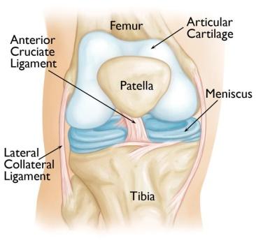
Your knee is the largest joint in your body and one of the most complex. The bones that make up the knee include the lower end of the femur (thighbone), the upper end of the tibia (shinbone), and the patella (kneecap).
Other important structures that make up the knee joint include:
- Articular cartilage. The ends of the femur and tibia, and the back of the patella are covered with articular cartilage. This slippery substance helps your knee bones glide smoothly across each other as you bend or straighten your leg.
- Synovium. The knee joint is surrounded by a thin lining called synovium. This lining releases a fluid that lubricates the cartilage and reduces friction during movement.
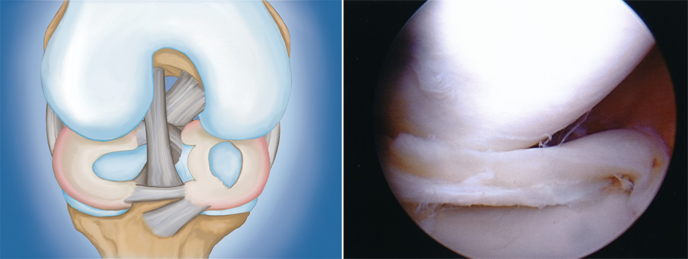
- Meniscus. Two wedge-shaped pieces of meniscal cartilage act as “shock absorbers” between your femur and tibia. Different from articular cartilage, the meniscus is tough and rubbery to help cushion and stabilize the joint.
- Ligaments. Bones are connected to other bones by ligaments. The four main ligaments in your knee act like strong ropes to hold the bones together and keep your knee stable.
- The two collateral ligaments are found on either side of your knee.
- The two cruciate ligaments are found inside your knee joint. They cross each other to form an “X” with the anterior cruciate ligament in front and the posterior cruciate ligament in back.
When Knee Arthroscopy is Recommended
We may recommend knee arthroscopy if you have a painful condition that does not respond to nonsurgical treatment. Nonsurgical treatment includes rest, physical therapy, and medications or injections that can reduce inflammation.
Knee arthroscopy may relieve painful symptoms of many problems that damage the cartilage surfaces and other soft tissues surrounding the joint.
Common arthroscopic procedures for the knee include:
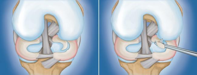
- Removal or repair of a torn meniscus
- Reconstruction of a torn anterior cruciate ligament
- Reconstruction of a torn posterior cruciate ligament
- Removal of inflamed synovial tissue
- Trimming of damaged articular cartilage
- Cartilage Transplant Techniques
- Removal of loose fragments of bone or cartilage
- Treatment of patella (kneecap) problems
- Treatment of knee sepsis (infection)
Preparing for Surgery
Anesthesia
Before your surgery, our anesthetist will examine you. Knee arthroscopy can be performed under local, regional, or general anesthesia:
- Local anesthesia numbs just your knee
- Regional anesthesia numbs you below the waist
- General anesthesia puts you to sleep
Surgical Procedure
Positioning
Once you are moved into the operating room, you will be given anesthesia. To help prevent surgical site infection, the skin on your knee will be cleaned. Your leg will be covered with surgical draping that exposes the prepared incision site.
At this point, a positioning device is sometimes placed on the leg to help stabilize the knee while the arthroscopic procedure takes place.
Procedure
To begin the procedure, the surgeon will make a few small incisions, called “portals,” in your knee. A sterile solution will be used to fill the knee joint and rinse away any cloudy fluid. This helps us see the structures inside your knee clearly and in great detail.
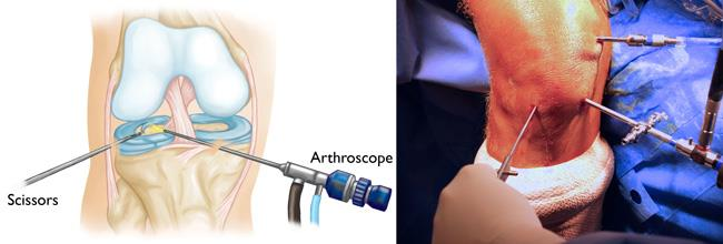
The first task is to properly diagnose your problem. Dr Kaushak will insert the arthroscope and use the image projected on the screen to guide it.
Specialized instruments are used for tasks like shaving, cutting, grasping, and meniscal repair. In many cases, special devices are used to anchor stitches into bone.
Closure
Most knee arthroscopy procedures last less than an hour or two. The length of the surgery will depend upon the findings and the treatment necessary.
We may close each incision with a stitch or steri-strips (small bandaids), and then cover your knee with a soft bandage.
Complications
The complication rate after arthroscopic surgery is very low. If complications occur, they are usually minor and are treated easily. Possible postoperative problems with knee arthroscopy include:
- Infection
- Blood clots
- Knee stiffness
- Accumulation of blood in the knee
Recovery
After surgery, you will be moved to the recovery room.
While recovery from knee arthroscopy is faster than recovery from traditional open knee surgery, it is important to follow your our team’s instructions carefully to make a full recovery.
Bearing Weight
Most patients need crutches or other assistance after arthroscopic surgery. We will tell you how and when it is safe to put weight on your foot and leg. Climbing and descending stairs with the walking aid will also be taught.
Rehabilitation Exercise
You should exercise your knee regularly for several weeks after surgery. This will restore motion and strengthen the muscles of your leg and knee.
Working with a physical therapist can help you achieve your best recovery.
Therapeutic exercise will play an important role in how well you recover. A formal physical therapy program will improve your final result.
Outcome
Many people return to full, unrestricted activities after arthroscopy. Your recovery will depend on the type of damage that was present in your knee.
Unless you have had a ligament reconstruction, you should be able to return to most physical activities after 6 to 8 weeks, or sometimes much sooner. Higher impact activities may need to be avoided for a longer time.
If your job involves heavy work, it may be longer before you can return to your job. Plaese discuss when you can safely return to work with Dr Kaushal.
For some people, lifestyle changes are necessary to protect the joint. An example might be changing from high impact exercise (such as running) to lower impact activities (such as swimming or cycling). These are decisions you will make after discussion with our team.
Sometimes, the damage to your knee can be severe enough that it cannot be completely reversed with surgery.
Wonderful, what a web site itt is! This webpage presenhts useful facts to us, keep
it up. https://odessaforum.biz.ua/