
Hip Arthroscopy
Hip arthroscopy is a surgical procedure that allows doctors to view the hip joint without making a large incision (cut) through the skin and other soft tissues. Arthroscopy is used to diagnose and treat a wide range of hip problems.
During hip arthroscopy, your surgeon inserts a small camera, called an arthroscope, into your hip joint. The camera displays pictures on a video monitor, and your surgeon uses these images to guide miniature surgical instruments.
Because the arthroscope and surgical instruments are thin, your surgeon can use very small incisions, rather than the larger incision needed for open surgery. This results in less pain for patients, less joint stiffness, and often shortens the time it takes to recover and return to favorite activities.
Hip arthroscopy has been performed for many years, but is not as common as knee or shoulder arthroscopy.
Anatomy
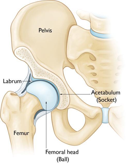
The hip is a ball-and-socket joint. The socket is formed by the acetabulum, which is part of the large pelvis bone. The ball is the femoral head, which is the upper end of the femur (thighbone).
A slippery tissue called articular cartilage covers the surface of the ball and the socket. It creates a smooth, frictionless surface that helps the bones glide easily across each other.
The acetabulum is ringed by strong fibrocartilage called the labrum. The labrum forms a gasket around the socket.
The joint is surrounded by bands of tissue called ligaments. They form a capsule that holds the joint together. The undersurface of the capsule is lined by a thin membrane called the synovium. It produces synovial fluid that lubricates the hip joint.
When Hip Arthroscopy Is Recommended
We will recommend hip arthroscopy if you have a painful condition that does not respond to nonsurgical treatment. Nonsurgical treatment includes rest, physical therapy, and medications or injections that can reduce inflammation.
Hip arthroscopy may relieve painful symptoms of many problems that damage the labrum, articular cartilage, or other soft tissues surrounding the joint. Although this damage can result from an injury, other orthopaedic conditions can lead to these problems, including:
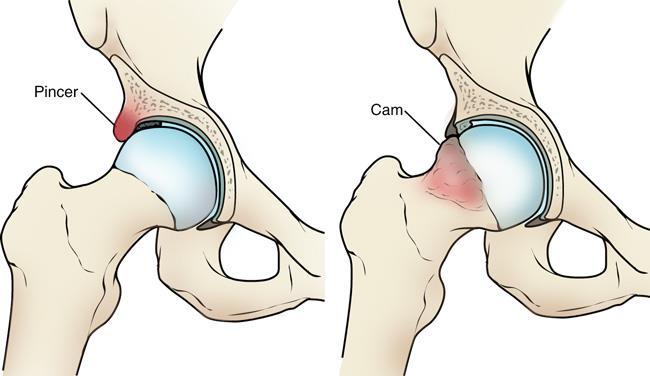
- Femoroacetabular impingement (FAI) is a disorder in which extra bone develops along the acetabulum (pincer impingement) or on the femoral head (cam impingement). This bone overgrowth—called spurs—damages the soft tissues of the hip during movement. Sometimes bone spurs develop in both the acetabulum and femoral head.
- Dysplasia is a condition in which the hip socket is abnormally shallow. This puts more stress on the labrum to keep the femoral head within the socket, and makes the labrum more susceptible to tearing.
- Snapping hip syndromes cause a tendon to rub across the outside of the joint. This type of snapping or popping is often harmless and does not need treatment. In some cases, however, the tendon is damaged from the repeated rubbing.
- Synovitis causes the tissues that surround the joint to become inflamed.
- Loose bodies are fragments of bone or cartilage that become loose and move around within the joint.
- Hip joint infection
Planning for Surgery
Anesthesia
Before the operation, you will also be evaluated by our anesthetist. Hip arthroscopy is most commonly performed under general anesthesia, where you go to sleep for the operation. Regional anesthesia, such as spinal or epidural, can also be used. With regional anesthesia, you are awake but your body is numb from the waist down.
Surgical Procedure
Positioning
At the start of the procedure, your leg will be put in traction. This means that your hip will be pulled away from the socket enough for Dr Kaushal to insert instruments, see the entire joint, and perform the treatments needed.
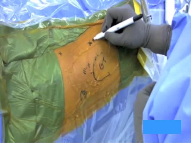
We will typically draw lines on the hip as a surface marking guide to indicate specific anatomy structures (such as bone, nerves, and blood vessels), as well as incision placements and portals for the arthroscope.
Procedure
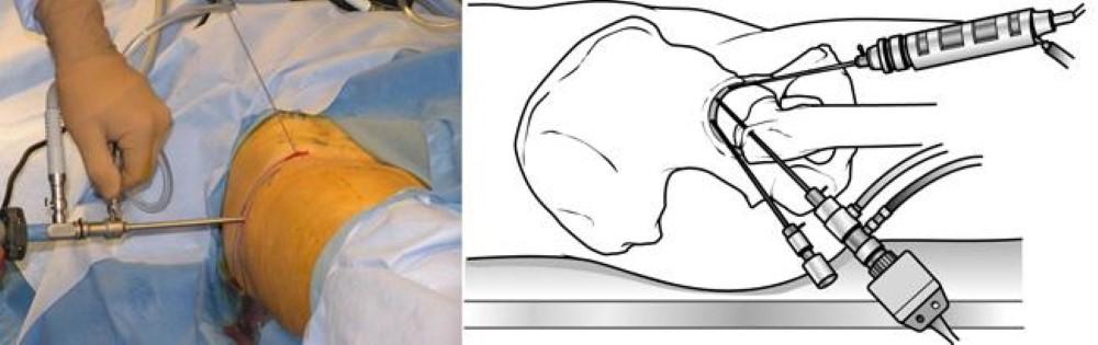
After traction is applied, we will make a small puncture in your hip (about the size of a buttonhole) for the arthroscope. Through the arthroscope, Dr Kaushal can view the inside of your hip and identify damage.
Fluid flows through the arthroscope to keep the view clear and control any bleeding. Images from the arthroscope are projected on the video screen showing your surgeon the inside of your hip and any problems. We will evaluate the joint before beginning any specific treatments.
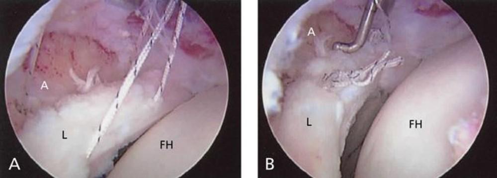
Once the problem is clearly identified, we insert other small instruments through separate incisions to repair it. A range of procedures can be done, depending on your needs. For example:
- Smooth off torn cartilage or repair it
- Trim bone spurs caused by FAI
- Remove inflamed synovial tissue
Specialized instruments are used for tasks like shaving, cutting, grasping, suture passing, and knot tying. In many cases, special devices are used to anchor stitches into bone.
The length of the procedure will depend on what problem we find inside the joint and the amount of work to be done. At the end of surgery, the arthroscopy incisions are usually stitched or covered with skin tapes. A padding dressing is applied.
Complications
Complications from hip arthroscopy are uncommon. Any surgery in the hip joint carries a small risk of injury to the surrounding nerves or blood vessels, or the joint itself. The traction needed for the procedure can stretch nerves and cause numbness, but this is usually temporary.
There are also small risks of infection, as well as blood clots forming in the legs (deep vein thrombosis).
Recovery
Bearing Weight
Crutches may be necessary after your procedure. In some cases, they are needed only until any limping has stopped. If you required a more extensive procedure, however, you may need crutches for 4-6 weeks.
We will develop a rehabilitation plan based on the surgical procedures you required. In most cases, physical therapy is necessary to achieve the best recovery. Specific exercises to restore your strength and mobility are important. Our therapist will also guide you with additional do’s and dont’s during your rehabilitation.
Long-Term Outcomes
Many people return to full, unrestricted activities after arthroscopy. Your recovery will depend on the type of damage that was present in your hip.
For some people, lifestyle changes are necessary to protect the joint. An example might be changing from high impact exercise (such as running) to lower impact activities (such as swimming or cycling). These are decisions you will make during discussion with our team.
Sometimes, the damage can be severe enough that it cannot be completely reversed and the procedure may not be successful.
Future Developments
Arthroscopy has helped physicians and researchers better understand many hip joint problems. As surgical techniques evolve, it is anticipated that hip arthroscopy will play a greater role in diagnosing and treating hip disease.