
A herniated disk is a condition that can occur anywhere along the spine, but most often occurs in the lower back. It is sometimes called a bulging, protruding, or ruptured disk. It is one of the most common causes of lower back pain, as well as leg pain or “sciatica.”
Between 60% and 80% of people will experience low back pain at some point their lives. Some of these people will have low back pain and leg pain caused by a herniated disk.
Although a herniated disk can be very painful, most people feel much better with just a few weeks or months of nonsurgical treatment.
Anatomy
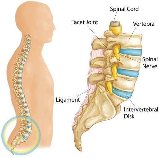
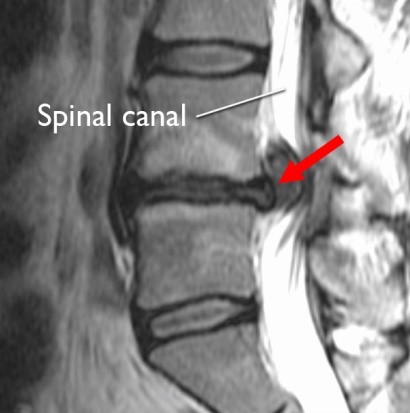
Your spine is made up of 24 bones, called vertebrae, that are stacked on top of one another. These bones connect to create a canal that protects the spinal cord.
Five vertebrae make up the lower back. This area is called your lumbar spine.
Other parts of your spine include:
Spinal cord and nerves. These “electrical cables” travel through the spinal canal carrying messages between your brain and muscles. Nerve roots branch out from the spinal cord through openings in the vertebrae.
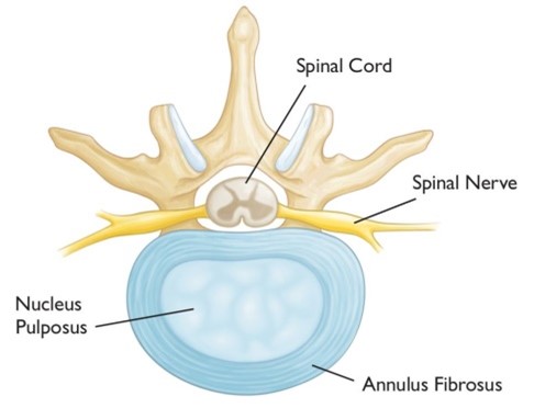
Intervertebral disks. In between your vertebrae are flexible intervertebral disks. These disks are flat and round, and about a half inch thick.
Intervertebral disks act as shock absorbers when you walk or run. They are made up of two components:
- Annulus fibrosus. This is the tough, flexible outer ring of the disk.
- Nucleus pulposus. This is the soft, jelly-like center of the disk.
Description
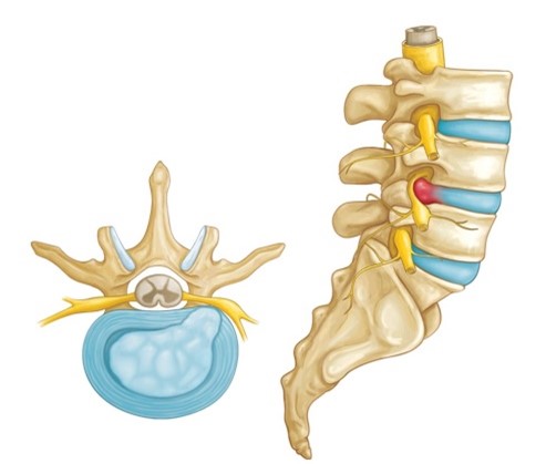
A disk begins to herniate when its jelly-like nucleus pushes against its outer ring due to wear and tear or a sudden injury. This pressure against the outer ring may cause lower back pain.
If the pressure continues, the jelly-like nucleus may push all the way through disk’s outer ring or cause the ring to bulge. This puts pressure on the spinal cord and nearby nerve roots. In addition, the disk material releases chemical irritants that contribute to nerve inflammation. When a nerve root is irritated, there may be pain, numbness, and weakness in one or both of your legs, a condition called “sciatica.”
Cause
A herniated disk is most often the result of natural, age-related wear and tear on the spine. This process is called disk degeneration. In children and young adults, disks have high water content. As people age, the water content in the disks decreases and the disks become less flexible. The disks begin to shrink and the spaces between the vertebrae get narrower. This normal aging process makes the disks more prone to herniation.
A traumatic event, such as a fall, can also cause a herniated disk.
Risk Factors
Certain factors may increase your risk of a herniated disk. These include:
Gender. Men between the ages of 20 and 50 are most likely to have a herniated disk.
Improper lifting. Using your back muscles instead of your legs to lift heavy objects can cause a herniated disk. Twisting while you lift can also make your back vulnerable. Lifting with your legs, not your back, may protect your spine.
Weight. Being overweight puts added stress on the disks in your lower back.
Repetitive activities that strain your spine. Many jobs are physically demanding. Some require constant lifting, pulling, bending, or twisting. Using safe lifting and movement techniques can help protect your back.
Frequent driving. Staying seated for long periods, plus the vibration from the car engine, can put pressure on your spine and disks.
Sedentary lifestyle. Regular exercise is important in preventing many medical conditions, including a herniated disk.
Smoking. It is believed that smoking lessens the oxygen supply to the disk and causes more rapid degeneration.
Symptoms
In most cases, low back pain is the first symptom of a herniated disk. This pain may last for a few days, then improve. Other symptoms may include:
- Sciatica. This is a sharp, often shooting pain that extends from the buttock down the back of one leg. It is caused by pressure on the spinal nerve.
- Numbness or a tingling sensation in the leg and/or foot
- Weakness in the leg and/or foot
- Loss of bladder or bowel control. This is extremely rare and may indicate a more serious problem called cauda equina syndrome. This condition is caused by the spinal nerve roots being compressed. It requires immediate surgical attention.
Medical History and Physical Examination
After discussing your symptoms and medical history, your doctor will perform a physical examination. The exam may include the following tests:
- Neurological examination. A neurological examination will help your doctor determine if you have any muscle weakness or loss of sensation. During the exam, he or she will:
- Check muscle strength in your lower leg by assessing how you walk on both your heels and toes. Muscle strength in other parts of your body may also be tested.
- Detect loss of sensation by checking whether you can feel a light touch on your leg and foot.
- Test your reflexes at the knee and ankle. These may sometimes be absent if there is a compressed nerve root in your spine.
- Straight leg raise (SLR) test. This test is a very accurate predictor of a disk herniation in patients under the age of 35. During the test, you lie on your back and your doctor carefully lifts your affected leg. Your knee stays straight. If you feel pain down your leg and below the knee, it is a strong indication that you have a herniated disk.
Imaging Studies
Magnetic resonance imaging (MRI) scan. These studies provide clear images of the body’s soft tissues, including intervertebral disks. Your doctor may order an MRI scan to help confirm the diagnosis and to learn more about which spinal nerves are affected.
If you are unable to tolerate an MRI, a computerized tomography (CT) scan, or a CT myelogram may be ordered instead.
Treatment
For the majority of patients, a herniated lumbar disk will slowly improve over a period of several days to weeks. Typically, most patients are free of symptoms by 3 to 4 months. However, some patients do experience episodes of pain during their recovery.
Nonsurgical Treatment
Initial treatment for a herniated disk is usually nonsurgical in nature. Treatment focuses on providing pain relief.
Nonsurgical treatment may include:
Rest. One to 2 days of bed rest will usually help relieve back and leg pain. Do not stay off your feet for longer, however. When you resume activity, try to do the following:
- Take rest breaks throughout the day, but avoid sitting for long periods.
- Make all your physical activity slow and controlled, especially bending forward and lifting.
- Change your daily activities to avoid movements that can cause further pain.
Nonsteroidal anti-inflammatory medications (NSAIDs). Medications such as ibuprofen or naproxen can help relieve pain.
Physical therapy. Specific exercises will help strengthen your lower back and abdominal muscles.
Epidural steroid injection. An injection of a cortisone-like drug into the space around the nerve may provide short-term pain relief by reducing inflammation.
Surgical Treatment
Only a small percentage of patients with lumbar disk herniation require surgery. Spine surgery is typically recommended only after a period of nonsurgical treatment has not relieved painful symptoms, or for patients who are experiencing the following symptoms:
- Muscle weakness
- Difficulty walking
- Loss of bladder or bowel control
Endoscopic Diskectomy. The procedure is done through a small incision at the level of the disk herniation and often involves the use of a small tube; through which an endoscope and instruments are passed under magnified vision.
The herniated part of the disk is removed along with any additional fragments that are putting pressure on the spinal nerve.
As there is no open surgery involved, the muscles and ligaments in the back are preserved leading to minimal post procedure back pain and early return to routine activities.
Rehabilitation. One is allowed to walk on the evening of the day of procedure. It is encouraged to use washroom to pass urine. Deep breathing exercises, active ankle, knee, hip movements are encouraged immediately.
To reduce the risk of repeat herniation, it is advisable to avoid forceful coughing, sneezing, constipation, repeated forward bending, twisting and lifting heavy weights for the first few weeks after surgery. The Annulus usually heals within 6-8 weeks post endoscopy after which restrictions are relaxed but patients are encouraged to improve their posture, strengthen their back and core muscles to prevent reherniation.
Considerations
With both surgical and nonsurgical treatment, there is a 5% to 10% chance that the disk will herniate again.
The risk of nonsurgical treatment is that your symptoms may take a long time to resolve. Patients who try nonsurgical treatment for too long before electing to have surgery may experience less improvement of pain and function than those who elect to have surgery earlier.
Surgical risks. There are minor risks associated with every surgical procedure. These include bleeding, infection, and reaction to anesthesia.
Specific complications from surgery for a herniated disk include:
Nerve injury
- Infection
- Tear of the sac covering the nerves (dural tear)
- Hematoma causing nerve compression
- Recurrent disk herniation
- Need for further surgery
Outcomes
Overall, the results of microdiskectomy surgery are generally very good. Patients tend to see more improvement of leg pain than back pain. Most patients are able to resume their normal activities after a period of recovery following surgery. Typically, the first symptom to improve is pain, followed by overall strength of the leg, and then sensation.